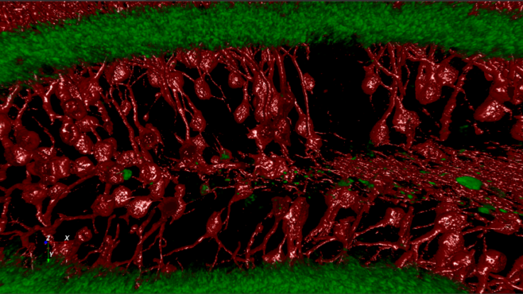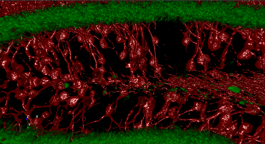

Mouse neuron network visualized in Aivia (Image credit: Margaret I Davis – NIAAA)
Aivia is revolutionizing academic and biomedical image analysis by democratizing powerful machine learning and virtual reality technologies.
— Luciano Lucas
BELLEVUE, WA, UNITED STATES, January 30, 2018 /EINPresswire.com/ — Aivia is revolutionizing academic and biomedical image analysis by democratizing powerful machine learning and virtual reality (VR) technologies. The tools now available in Aivia can accelerate research into treatments for Alzheimer’s, Parkinson’s, dementia, ALS and many other brain related conditions.
Machine learning classification algorithms have the potential to both minimize human biases and drastically increase the productivity both in a pharma/drug discovery setting and in academic research. First, Aivia captures the input provided by a domain expert while he reviews an image and shows it examples of each phenotype of interest. Second, the software autonomously extracts and categorizes numerous hidden features and in doing so, learns to identify the different phenotypes. Finally, the trained software can be used on other images resulting in the (automatic) detection of cell phenotypes of interest – a key procedure when learning about the healthy brain or when testing novel drugs.
Aivia is also pioneering the use of VR for the exploration of 3D/4D images. This visualization modality offers the most natural and insightful experience currently in existence. The user is immersed in the 3D/4D data set and has the freedom to walk around, look in any direction and intuitively interact with the shown objects (e.g. neurons and spines). This type of visualization often enables neuroscientists to see cellular relationships they had never seen before thus expanding our general understanding of neuronal structure and function.
“Aivia combines the most advanced VR technology with powerful, yet easy to use, phenotype detection. Moreover, it also includes a wide range of image analysis tools such as fully featured automated neuron and spine detection. Aivia is currently the only software that combines these three powerful technologies thus enabling unprecedented levels of productivity and insight. We believe we have created a toolbox that will positively impact the rate of discovery in the field of neurosciences” says Luciano Lucas (PhD), the EVP at DRVISION.
A human brain is thought to be composed of 8 to 20 cell types and more than 86 billion neuronal cells. The morphology and spatial distribution of these cells follows a set pattern in healthy individuals. This pattern is characteristically altered in the brain of people affected by neurodegenerative diseases and other neurological disorders. Until now an expert would manually detect and classify each imaged cell into a specific phenotype – a process that can take hours for a single data set – researchers typically analyze dozens to hundreds of such data sets per study. With Aivia the same process takes less than 1 minute and is not affected by human errors related to fatigue and some types of biases.
An United Nations report shows that 1 in 6 humans suffer from at least one neurological disorder in their lifetime, that is over 1 billion people worldwide – moreover, 6.8 million people died in 2016 as a result of these types of conditions. The annual cost of healthcare for patients affected by neuropathologies, in Europe alone, is over 139 billion Euro. Moreover, the brain is the least understood organ in the human body. This fact has recently fueled the start of several international efforts to study brain cellular structure and function (e.g. BRAIN Initiative, Human Connectome Project). Aivia’s innovative functionality is poised to help neuroscientists answer questions faster and with fewer biases.
“We are committed to creating tools that enable the advancement of scientific discovery. Aivia is the leading tool in a new generation of software that leverages fast machine learning (ML). Aivia will gradually gain additional novel fast ML and deep learning algorithms for both classification and image segmentation. A new era is starting in the field of microscopy image processing.” says Luciano Lucas (PhD) the EVP at DRVISION.
About DRVISION Technologies
DRVISION Technologies LLC was founded by Dr. James Shih-Jong Lee in 1999 (as SVision) who was the CTO and co-founder of NeoPath – the company that created the FDA approved automated pap smear system – BD FocalPoint GS (now part of BD Diagnostics). DRVISION holds 50 US patents in machine learning, pattern recognition, particle tracking and other image processing technologies. DRVISION has developed numerous applications for other companies in a wide range of markets (biomedical research, biotech, pharma, pathology, industrial manufacturing, quality control, etc.) and covering numerous applications.
Quyen Tran
DRVISION Technologies
1-855-423-5577
email us here
![]()
This Article Was Originally Posted at www.einnews.com